Basic HTML Version


WHEN
healthcare providers
need to know what’s going on inside
your body, there’s a wide range of
sophisticated imaging devices that
can help them find out. By using this
equipment, your medical team can
get images of bones, organs, muscles,
blood vessels and cartilage—all with-
out having to do surgery.
This guide from Benjamin Seckler,
MD, Chief of Radiology, and other
experts explains some of the most
current imaging techniques used.
X-ray
What it is:
Medical x-rays are
used to detect disease or injury early
enough for a medical problem to be
managed, treated or cured. X-rays
provide a two-dimensional image of
the body’s tissues.
X-rays are the most common
and widely available imaging tech-
nique. During an x-ray, electromag-
netic radiation waves form images
of structures inside the body. As
x-rays penetrate the body, they are
absorbed in different amounts by dif-
ferent body tissues. For instance, bones
are dense and absorb x-rays very well,
but soft tissues (skin, fat and muscle)
allowmore x-rays to pass through. The
result is an image in which bones ap-
pear white and soft tissues appear gray.
Even structures not normally vis-
ible on x-rays, such as blood vessels,
can be seen after a substance—known
as a contrast medium—is injected,
swallowed or given as an enema.
What it’s used for:
X-rays are very
versatile—providers use them for
everything from checking for broken
bones to looking for cancer. Low-
dose x-rays examine soft tissues, such
as the breast, and are widely used to
screen for breast cancer.
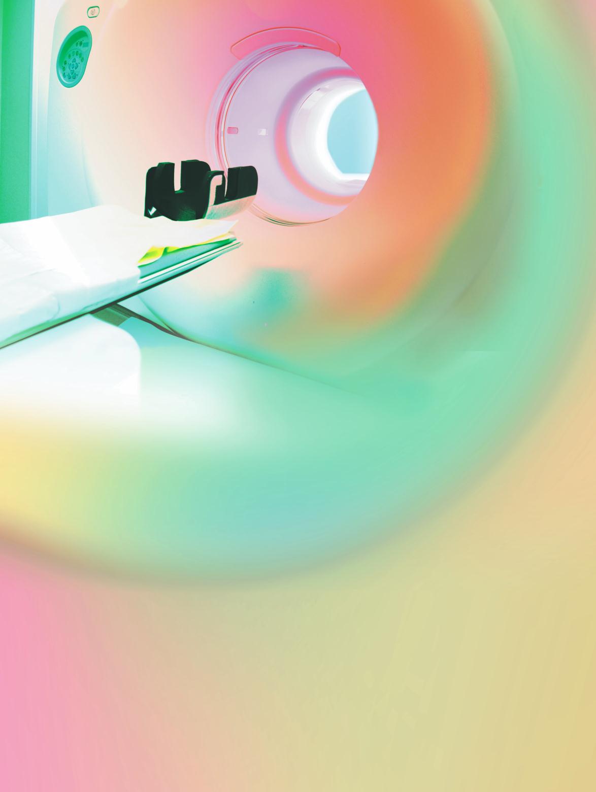
Computed tomography (CT)
What it is:
CT uses special x-ray
equipment to take images from vari-
ous angles around the body. Comput-
ers then process the x-rays, creating a
detailed cross-sectional view of body
tissues and organs. CT scans provide
X-ray
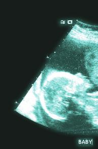
Ultrasound
Computed tomography (CT)
IMAGING
4
q
lifeand health
Sharon Hospital is a Breast
Imaging Center of Excellence
and holds the ACR Gold
Standard of Accreditation.